This is a video of Dr. Kate von Klemperer discussing surveillance of AD patients.
Transcript (auto-generated)
This transcript was generated automatically and may contain errors.
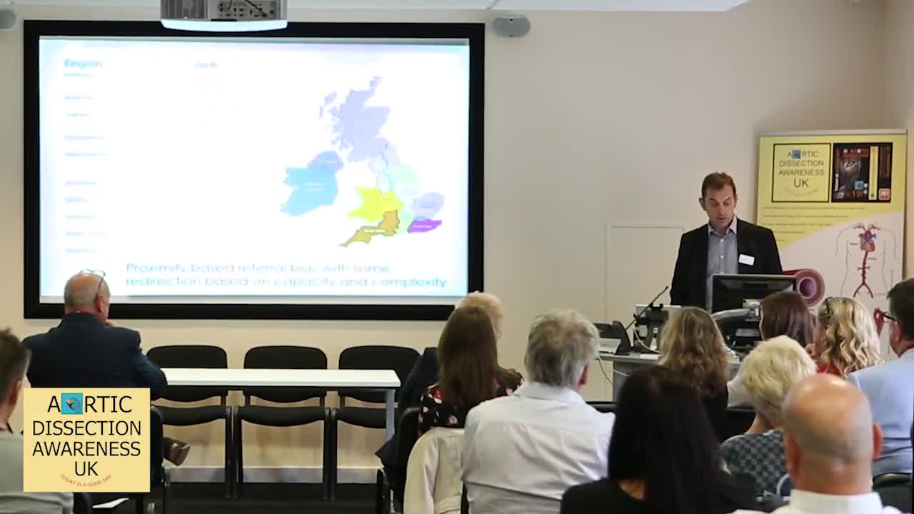
So Kate von Kemper is going to come and tell us about what she does with BGEL and she's going to talk specifically about those patients who we're doing surveillance on. It might be a little bit of overlap here, as Bijal and I do, and Elena do very similar jobs. But I'm focusing on surveillance today. And I don't have to tell this room how devastating dissection can be for patients and unfortunately the aneurysmal disease that often precedes dissection is an indolent disease and patients are often asymptomatic with it and it's really important to be able to identify patients at risk because at the moment the only sort of robust management plan we have for patients to prevent dissection is replacement of the aneurysmal aorta. So B-GELs sort of describe the aorta to you. The aorta pumps about 200 million liters of blood in our lifetime, and for our purposes we divide the aorta into the ascending aorta, the arch, which you can see there, and then and the descending aorta, and aneurysms and dissections of the aorta are classified according to those sections. And as Sandy touched on, in medicine, we've all got very specialized. And actually, the medical teams dealing with each portion of the aorta are different as well. And therefore, this, again, is a common theme, and it'll keep being the theme of today. We require multidisciplinary care in looking after these patients. And in our aortovascular service, we are referred all comers with aortic disease. And after an initial assessment, which Bijal's described to you, taking a good family history, doing a clinical assessment. We need to ask ourselves a few things. The first is, what is normal? So particularly in patients that we're screening for aortic disease, we need to understand what we would consider normal in the person sitting in front of us. We need to know what the best method of investigating that person's aortovascular system is. And that would very much be based upon our initial clinical assessment. And then in those patients who have aortic disease, we need to know the best way to follow them up and at what intervals to do so. And very importantly, we need to know when to intervene. So this looks like quite a complicated graph system here, but it's actually quite straightforward. These are normal aortic dimensions men on the left-hand side and women on the right-hand side, and already you can see here that actually if you're six foot tall as a man, we'd expect you to have a bigger aortic diameter than if you're six foot tall as a woman. So it's gender specific, and you can also see here that if you're taller we expect you to have a bigger aortic diameter than someone who's four foot. And that as Bijal said, the aorta increases in size with age as the aorta loses its elasticity. The aortic, the increase in size is approximately one millimeter over decades, so it's really small increases with age. And we know that in our patients with bicuspid valve, that increase in size with age is quicker, so they tend to increase by five millimeters over a decade. And in some of the patients with connective tissue disease, and in particular the patients with Marfan and familial thoracic aortic disease, we see a much quicker progression in the size of the aorta and that guides us on how we follow patients up because we don't want to create anxiety in patients with normal sized aortas where we keep bringing them back to clinic on a yearly basis and we might increase their interval if we just want to keep an eye on them. So does size matter and as Bijal again has shown there was some good retrospective data in the 1990s which showed a significant increase in your risk of dissection as your aortic size increased, and this is from the Yale group. And that guided those initial sort of ballpark guidelines that you should be replacing ascending aortas at 5.5 centimeters. You'll see in this graph that dissections do happen at smaller sizes as well, but when we look back at that data and we look at the population risk, your relative risk of having a dissection at a bigger aorta is significantly higher than at a smaller aorta. However, that group of patients who have normal lay orators are one of our challenges for future research. So I'm not sure if you can see that, but in terms of our ongoing surveillance and deciding on how to investigate and follow patients, we need to have a method that's accessible. We need to have a standardized way of measuring. We're talking millimeters here, so we need to be able to put images up next to each other and be quite confident that there's been significant growth. And we need a safe and cost-effective method. And it needs to be appropriate to the condition that the patient has. So this, I'm sure all of the patients in the room will certainly be familiar with, is an echocardiogram. It's traditionally how we have followed up patients. And a lot of the literature on aortic disease is based on echo measurements. So you can see here, for those of you who are not familiar with it, this is the aorta valve here. This is the base of the aorta, the sinus valve there. And this is the opening aorta, which comes out of the left ventricle, the heart lying on its left-hand side. And you can see here how we make our measurements. And whilst this is a very good method of following up the base of the aorta, which can be useful for patients with a myofan type of phenotype, where that's the area that tends to be dilated, it's not as good a method for following up the rest of the aorta. And you can see here the caliber of images that we get of the rest of the aorta. And it's certainly not good enough for following up patients who've had previous dissection. So we tend to use it as a surveillance tool that's readily available to all of us, but often we need other ways of imaging the aorta. And this moves us on to a CT scan, which, again, is quick and accessible to us in our hospitals, gives us a quick, easy answer, which is very reproducible, gives us some beautiful 3D imaging. And we can assess all the complications related to dissection with this. And so a lot of our patients who've had dissection will have relatively regular CTs, certainly in the first few years after dissection. But the sort of limitations or the downfall of CT scan is that it requires contrast, which can affect the kidneys. So if people have got kidney problems, it's an issue. And it's also, as we all know, it exposes us to radiation, which scan by scan is not an issue. But certainly in our younger patients, who B-gel and I see lot of, you wouldn't want to be doing it long-term. So CT scan, if necessary, provides us with excellent information, but as a long-term surveillance tool, we tend to use MRI scans. And I'm afraid, after all these beautiful pictures, this is not going to play for me today, but this is just some imaging from Christian Mortenson, one of our imaging consultants, and he's got a beautiful video just showing how they line up their aorta to make sure that they're getting the dimensions correct with MRI scanning. An MRI gives us a lot of extra information. It gives us information about the valves. It gives us information about the heart function. It gives us all the same information about adjacent structures, And it can give us some very beautiful 3D angiograms as well. So as Bijal showed, aneurysms can develop in different areas, and there's certain – we know that there's certain patterns of vascular involvement with various conditions. And so we will tailor the tests that we do to that. You can see here the tortuous vessels on the right of Lois Dietz, as Bijal showed, and the Marfan phenotype, or Lois Dietz phenotype, of root in the middle, compared to the bacuspid valve on the right, the far right. In our vascular EDS patients, who we know have a medium vessel as well as aortic involvement, We need a way of screening for aneurysms elsewhere. These are patients I see with Paul Flora in my clinic because they often do have problems which relate to their small vessels, not only their aorta. And this is the protocol that our imaging team have developed, which is basically a surveillance screen from the neck down to the pelvis. And so it gives us all of the information that we need. And to guide us, as mentioned earlier, we've got some good guidance based on the current literature on when we should be intervening. And why intervene? Well, we know that emergency surgery has a much higher risk and much worse outcomes long-term than elective surgery in this setting. And this is the data from John-Olef Teriades, one of the sort of forefathers of aortic surgery, and he's got his, he presents his 30-year experience of prophylactic valve and root replacement, and this sort of shows very beautifully So this, you can see there's a light gray line there, which is normal and expected survival. And you can see his line of his patients who've had a valve and root replacement, and their survival is very similar. So it's a good take-home message, and we certainly get very good results, and we know that intervening is important. and worthwhile. I think, sort of, going forward in terms of our imaging and surveillance, as I said, there's a whole group of people that we find difficult to assess risk in. We are unable at the moment to assess the strain on the aorta, and there's a lot of room for future research into why people dissect at smaller diameters and lots of work to be done. The work, the research work done on aortas needs to be a life project like John Elefteriades has had because it takes a long time to get outcome data on aortic disease and that's why it's so great to be part of a team like we have here of people who are passionate about aortas and to have this sort of lifelong experience from pediatric through to dissection and through to old age. Thank you very much. you

 Health Shared



Comments