Health Shared Logo whiteHealth Shared Logo dark

Carpal Tunnel Decompression - Orthopaedic Surgery

Mr David WheelerMr David Wheeler

Surgical treatment of carpal tunnel syndrome involves cutting the transverse carpal ligament to release compression of the median nerve. The procedure is done under local anaesthetic and takes around 10-20 minutes. Most patients will be able to go home on the day of their operation. Most patients are helped by these procedures and will be able to return to their jobs and daily activities successfully with no long-term problems.

To begin, the patient lies flat on an operating table with their hand on a hand table with the hand and forearm numbed by local anaesthetic. The surgeon will test if pain can be felt; if so then more anaesthetic is given, or if not, then the procedure can begin. If the patient is overly anxious they can request a sedative to help keep calm.

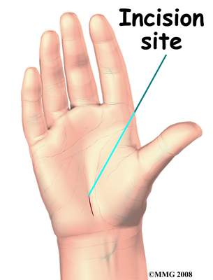
To help visualise the procedure, an initial 1-2 inch incision is made, the location of which depends on the type of surgery the surgeon has chosen. In open surgery, the surgeon separates the skin and works through an "open" incision to cut the transverse carpal ligament. In endoscopic surgery, the surgeon makes an incision to insert an endoscope, a small camera, into the incision and then cuts the ligament. These are shown below.

Related imageRelated image

Following the initial incision, subcutaneous fat and palmar tissue, fat below the skin and sometimes some palm muscle, is dissected to expose the transverse carpal ligament, the band of tissue responsible for the compression and pain. The ligament is then separated from the nerves and blood vessels underneath it and cut to release the median nerve. Once the nerve has been checked to make sure there is no more compression, the incision is closed and stitched up.

Once the procedure has been completed, the hand and forearm will still be numb and care should be taken to avoid using or hitting anything with it. The doctor will prescribe pain medication following the operation and the patient will usually be allowed to go home if there are no complications. The patient will be scheduled for a follow-up 2 weeks after the operation in an outpatient setting and then again at 3 months after to assess progress. Recovery is usually 6-12 weeks for full return to previous activities. Usually, the surgeon will be able to provide their own estimate upon request.


More from this author:

Mr David WheelerMr David Wheeler

Anterior Cruciate Ligament (ACL) Reconstruction - Orthopaedic Surgery

The Anterior Cruciate Ligament (ACL) is one of two crossing ligaments, along with the Posterior Cruciate Ligament (PCL) , found inside the knee which connect the femur (thighbone) and the tibia (shinbone) . These cruciate ligaments control back and forth movement of the lower leg and act to hold the bones together as well as stabilise the whole knee. Ligament injuries are considered "sprains" and can be graded by severity; grade 1 sprains - the ligament has been slightly stretched and becomes mildly damaged but can still keep the knee joint stable, grade 2 sprains (sometimes referred to as a partial tear) - the ligament is stretched until it becomes loose, and grade 3 sprains (often referred to as a complete tear ) - the ligament has been stretched until it splits into two pieces. The ACL can be injured in multiple ways, usually through sudden powerful movements which put too much stress on the knee. These include changing direction rapidly, stopping suddenly, slowing down while running, landing from a jump incorrectly, and direct contact or collision on the knee which can overextend the lower leg . These injuries can be prevented by strengthening of the leg muscles which support the load of the ligaments, strengthening of the core muscles, and training for correct technique in movements that place stress on the knee. Before treatment is discussed, a physical exam is used to assess for swelling and tenderness as well as the range of motion and overall function of the knee. Further techniques such as X-rays, to check for a bone fracture, and MRI or Ultrasound scans, to check for signs of damage in the tissues of the knee, are used to clarify the severity and location of the damage. Treatment for ACL injuries depends on the severity . Slight sprains or partial tears can be treated conservatively with rest, ice application, compression, and elevation (as known as the RICE steps ) as well as potentially a brace and physiotherapy to strengthen the supporting leg and core muscles. Complete tear treatment depends on the patient; if they are elderly or have a very low activity level then surgery may not be needed and the previous treatments can be used. However, if the patient wants to return to sports or activities that involve movements particularly stressful to the knee, such as basketball, football or rugby, then surgery is the only option remaining .