Health Shared Logo whiteHealth Shared Logo dark

Carotid endarterectomy for asymptomatic patients

prof. Usman Jafferprof. Usman Jaffer

Why is it done?

The procedure is done to remove an area of narrowing in the Internal Carotid artery which supplied the brain. This area of narrowing is called an atheromatous plaque. The purposed of the procedure is to prevent small pieces of this plaque/ narrowing from breaking off and travelling up to lodge in the brain (embolisation). This embolisation can cause stroke or TIA (mini-stroke)

The Intended benefit is to reduce the stroke.

The benefit is the reduction in risk of ipsilateral stroke. The Asymptomatic Carotid Surgery Trial (ACST) and Asymptomatic Carotid Atherosclerosis Study (ACAS) reported a net reduction in stroke risk of 5.4% and 5.9% respectively over medical therapy over 5 years (1, 2). The ACAS and especially the ACST trials showed that the net benefit of carotid endarterectomy (CEA) is delayed, as the asymptomatic population has an overall worse outcome after CEA for many months up to 2 years because of the perioperative morbidity (2). The estimated risk of ipsilateral stroke in asymptomatic carotid atherosclerosis with stenosis more than 50% is approximately 0.5 to 1% annually (3). Multispecialty guidelines states that carotid endarterectomy should be considered for asymptomatic patients who have more than 70% stenosis of internal carotid artery if the risk of perioperative stroke, myocardial infarction and death is low (less than 3%) (4, 5).

Will I need to do any preparation?

Generally you will need to fast (not eat or drink) before the procedure. This is usually six hours for food and two hours for clear liquids. Taking blood thinning medicine is important to reduce the risk of clot building up on the rough area to be removed from the carotid artery.

Anaesthesia

The procedure can be done under general or local anaesthetic. Some surgeons prefer local anaesthesia because it allows direct monitoring of your brain function. If you are awake, you can be asked to talk to us or squeeze a ball in order to tell that your brain is receiving adequate blood supply (perfusion). Although a study has been performed which shows no significant difference in outcome from surgery performed under local or general anaesthetic, some surgeons prefer to perform the procedure under local for the ability to minimise the use of carotid shunting (see later).

What does it involve?

The procure involves a number of steps, not all necessarily need to be done depending on your individual situation.

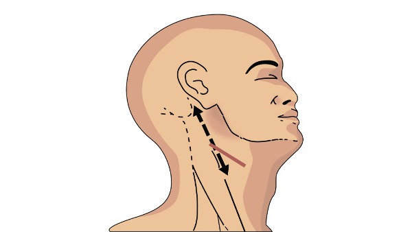
Firstly, you need to be comfortably positioned on the operating table. You will asked to look over to the opposite side to that being operated on to allow access to the neck.

The skin incision (cut) can be more vertical or more horizontal depending on the preferences of the surgeon. A more vertical cut allow better access to the structures being operated on whereas a more horizontal one tends to heal with a better scar.


Scar line for Carotid Endarterectomy. The black line provides better access whereas the brown more horizontal line has better cosmetic outcome.

The next step involves freeing the Carotid arteries from the surrounding tissue. The main artery passing through the neck is called the Common Carotid Artery. This splits into two arteries (Internal and External Carotid arteries). The Internal Carotid artery supplies the brain and the External carotid artery supplied the side of the head, All three need to be freed up.


Diagram of the Internal Jugular Vein in front of the Carotid arteries. The vertical yellow structure between the artery and vein is the Vagus nerve. The horizontal yellow structure in from of the arteries is the hypoglossal nerve.

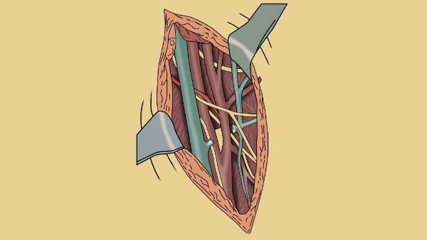
Care must be taken to free up the structures without damaging important nerves. The Vagus nerve lies between the artery and the vein. Damage to the Vagus nerve can cause a horse voice - this is usually temporary. The hypoglossal nerve above travels horizontally over the Internal and External carotid arteries. Damage to this cause weakness to one side of the tongue. This will cause the tongue to deviate to the damaged side when sticking it out. This is usually temporary also.

Once the arteries are freed up, heparin medicine is given to prevent blood clots building up in the blood when the arteries are clamped off. The arteries are clamped off and the diseased segment is opened. The narrowed area is cleaned - a process called endarterectomy.


Yellow atheroma material being removed from a diseased Internal Carotid artery.

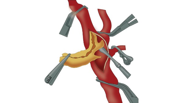
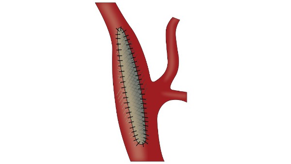
Once all the atheroma is removed and the inside of the artery is adequately cleaned, the artery is closed. This can be done directly with stitches or with a patch repair. It is important not to narrow the Internal Carotid artery when closing it - the patch can help with this.


Diagram of a close Carotid artery with a patch repair in place.

How long does it take? 

The procedure can take up to two hours normally. There will be additional time take for anaesthetic preparation and usually time spent after the operation in the recovery area.

Post procedure course (follow-up)

A drain may be attached to the neck to remove any fluid or blood that build up around the operation site for the first 24 hours. You may well be treated in the High Dependancy unit for the first 12-24 hours after surgery. This is done so your blood pressure can be accurately monitored using a monitoring device inserted into the radial artery at your wrist. If the BP strays high or low then this can be accurately treated which is important for the first 12 hours after surgery.

How long will I stay in hospital?

People often go home the next day following surgery.

What care will I need at home?

No particular care requirements are needed. The wound should generally be kept dry for around five days following the procedure and covered with a dressing. Whichever tablets your doctors have prescribed should be taken.

Will I need someone to stay with me?

No particular need, you should. be fully independant.

Will I need any special equipment when I go home?

No particular requirements.

What follow up care is needed? 

Often a follow up Duplex ultrasound scan is performed to ensure no technical issues exist in the blood flow through the carotid artery.

When can I start my normal activities again (e.g. driving, sports or work?)

Normal activities can be started immediately. Driving should be discussed with your stroke physician as there may be specific restrictions on your activity following a stroke or mini-stroke.


More from this author:

prof. Usman Jafferprof. Usman Jaffer

Carotid endarterectomy for symptomatic patients

prof. Usman Jafferprof. Usman Jaffer

Femoral-Below knee Popliteal Artery Bypass Graft

prof. Usman Jafferprof. Usman Jaffer

Endo-Vascular Infra-Renal Abdominal Aortic Aneurysm repair (EVAR)

prof. Usman Jafferprof. Usman Jaffer

Femoral- Above the knee Popliteal Bypass Graft

prof. Usman Jafferprof. Usman Jaffer

What are the symptoms of an Abdominal Aortic Aneurysm?

prof. Usman Jafferprof. Usman Jaffer

Anal Fistula

Treatment of anal fistula: A fistula without symptoms found on regular check up usually requires no therapy . [3,4] In case of anorectal abscess surgery for fistula repair should not be performed (unless the fistula is superficial and the tract is obvious). In the acute phase, simple incision and drainage of the abscess are sufficient. [5] Only 7-40% of patients will develop a fistula. Recurrent anal sepsis and fistula formation are twofold higher after an abscess in patients younger than 40 years and are almost threefold higher in non diabetics. Preoperative considerations include the following: • Rectal irrigation with enemas should be performed on the morning of the operation • Anesthesia can be general, local with intravenous sedation, or a regional block • Administer preoperative antibiotics Intraoperative considerations include the following: • Examine the patient under anesthesia to confirm the extent of the fistula • Identifying the internal opening (inside the anus) to prevent recurrence is imperative • A local anesthetic block at the end of the procedure provides postoperative analgesia Fistulotomy: The laying-open technique (fistulotomy) is useful for 85-95% of primary fistulas (ie, submucosal, intersphincteric, and low transsphincteric). [6, 7, 8, 9] The most common type of surgery for anal fistulas is a fistulotomy. This involves cutting along the whole length of the fistula to open it up so it heals as a flat scar.A fistulotomy is the most effective treatment for many anal fistulas, although it's usually only suitable for fistulas that don't pass through much of the sphincter muscles, as the risk of incontinence is lowest in these cases. If your surgeon has to cut a small portion of anal sphincter muscle during the procedure, they will make every attempt to reduce the risk of incontinence.In cases where the risk of incontinence is considered too high, one of the procedures below may be recommended instead. Seton techniques: If your fistula passes through a significant portion of anal sphincter muscle, your surgeon may initially recommend inserting a seton. A seton is a piece of surgical thread that is left in the fistula for several weeks to keep it open. This allows it to drain and helps it heal, while avoiding the need to cut the sphincter muscles. Loose setons allow fistulas to drain, but don't cure them. To cure a fistula, tighter setons may be used to cut through the fistula slowly. This may require several procedures that your surgeon can discuss with you. Seton Placement: A seton can be placed alone, combined with fistulotomy, or in a staged fashion. This technique is useful in patients with the following conditions [10, 11, 12] : • Complex fistulas (ie, high transsphincteric, suprasphincteric, extrasphincteric) or multiple fistulas • Recurrent fistulas after previous fistulotomy • Anterior fistulas in female patients • Poor preoperative sphincter pressures • Patients with Crohn disease or patients who are immunosuppressed Single-stage seton (cutting): Pass the seton through the fistula tract around the deep external sphincter after opening the skin, subcutaneous tissue, internal sphincter muscle, and subcutaneous external sphincter muscle. The seton is tightened down and secured with a separate silk tie. With time, fibrosis occurs above the seton as it gradually cuts through the sphincter muscles and essentially exteriorizes the tract. The seton is tightened on subsequent office visits until it is pulled through over 6-8 weeks. A cutting seton can also be used without associated fistulotomy. Recurrence and incontinence are important factors to consider when this technique is employed. The success rates for cutting setons range from 82-100%; however, long-term incontinence rates can exceed 30%. [13,14, 15] Two-stage seton (draining/fibrosing): Pass the seton around the deep portion of the external sphincter after opening the skin, subcutaneous tissue, internal sphincter muscle, and subcutaneous external sphincter muscle. Unlike the cutting seton, the seton is left loose to drain the intersphincteric space and to promote fibrosis in the deep sphincter muscle. Once the superficial wound is healed completely (2-3 months later), the seton-bound sphincter muscle is divided. Two studies (74 patients combined) supported the two-stage approach with a 0-nylon seton. Once wound healing is complete, the seton is removed without division of the remaining encircled deep external sphincter muscle. The researchers reported eradication of the fistula tract in 60-78% of cases. Mucosal Advancement Flap: A mucosal advancement flap is reserved for use in patients with chronic high fistula but is indicated for the same disease process as seton use. [1,16, 17] Advantages include a one-stage procedure with no additional sphincter damage. A disadvantage is poor success in patients with Crohn disease or acute infection. This procedure involves total fistulectomy, with removal of the primary and secondary tracts and completes excision of the internal opening. A rectal mucomuscular flap with a wide proximal base (two times the apex width) is raised. The internal muscle defect is closed with an absorbable suture, and the flap is sewn down over the internal opening so that its suture line does not overlap the muscular repair. Plugs and Adhesives: Advances in biotechnology have led to the development of many new tissue adhesives and biomaterials formed as fistula plugs. By their less-invasive nature, these therapies lead to decreased postoperative morbidity and risk of incontinence, but long-term data are lacking for eradication of disease, especially in complex fistulas, which carry high recurrence rates. [18, 19, 17] Reported series exist of fibrin glue treatment of fistula-in-ano, with 1-year follow-up showing recurrence rates approaching 40-80%. [20, 21,22] The Surgisis fistula plug has also had mixed long-term results in direct clinical trials. [23, 24, 25] Early success rates have been reported for newer materials, such as acellular dermal matrix and the bioabsorbable Gore Bio-A fistula plug, in low fistulas and good animal model data. [26] Assessment of long-term success rates with plug techniques for complex disease will be based on further data from randomized trials. In a randomized, controlled study designed to evaluate the efficacy and safety of the anal fistula plug in patients with fistulizing anoperineal Crohn disease, Senéjoux et al did not find the plug to be superior to seton removal for achieving fistual closure, regardless of whether the fistula was simple or complex. [27] A combined sphincter-sparing repair that includes both an anal fistula plug and a rectal advancement flap has been proposed for the treatment of transsphincteric fistula-in-ano. [28] LIFT Procedure: Ligation of the intersphincteric fistula tract (LIFT) is a sphincter-sparing procedure for complex transsphincteric fistulas first described in 2007. It is performed by accessing the intersphincteric plane with the goal of performing a secure closure of the internal opening and by removing the infected cryptoglandular tissue. [29] The intersphincteric tract is identified and isolated by performing meticulous dissection through the intersphincteric plane after making a small incision overlying the probe connecting the external and internal openings. Once isolated, the intersphincteric tract is hooked with a small right-angle clamp, and the tract is ligated close to the internal sphincter and then divided distal to the point of ligation. Hydrogen peroxide is injected through the external opening to confirm the division of the correct tract. The external opening and the remnant fistulous tract are curetted to the level of the proximity of the external sphincter complex. Finally, the intersphincteric incision is loosely reapproximated with an absorbable suture. The curettaged wound is left opened for dressing.[29,30,31] Because of its relative novelty, LIFT has not been extensively researched. In a randomized trial of 39 patients with complex fistula-in-ano who had failed previous procedures and were treated with LIFT technique, success rates were comparable to those seen with the anorectal advancement flap technique. [32] The probability of recurrence at 19 months was 8% for the LIFT technique versus 7% for the anorectal advancement flap. Time to return to work was shorter in the LIFT group (1 vs 2 wk), but there was no difference in incontinence scores. [32] Further randomized surgical trials are needed to determine whether this technique is a viable—or, possibly, a better—alternative to the other previously mentioned procedures for the treatment of fistula-in-ano. Diversion: In rare cases, the creation of a diverting stoma may be indicated to facilitate the treatment of a complex persistent fistula-in-ano. The most common indications include, but are not limited to, patients with perineal necrotizing fasciitis, severe anorectal Crohn disease, reoperative rectovaginal fistulas, and radiation-induced fistulas. Fecal diversion alone is effective in these select patients to control sepsis and symptoms; however, long-term success rates after reanastomosis are low because of recurrence from the underlying disease. Thus, this approach should be avoided unless the underlying fistula-in-ano disease process is repaired or has healed completely, which is unlikely. Postoperative Care: After the operation, most patients can be treated in an ambulatory setting with discharge instructions and close follow-up care. Sitz baths, analgesics, and stool-bulking agents (eg, bran and psyllium products) are used in follow-up care. Complications: Early postoperative complications may include the following: • Urinary retention • Bleeding • Fecal impaction • Thrombosed hemorrhoids Delayed postoperative complications may include the following: • Recurrence • Incontinence (stool) • Anal stenosis - The healing process causes fibrosis of the anal canal; bulking agents for stool help to prevent narrowing • Delayed wound healing - Complete healing occurs by 12 weeks unless an underlying disease process is present (ie, recurrence, Crohn disease) Postoperative rates of recurrence and incontinence vary according to the procedure performed, as follows: • Standard fistulotomy - The reported rate of recurrence is 0-18%, and the rate of any stool incontinence is 3-7% • Seton use - The reported rate of recurrence is 0-17%, and the rate of any incontinence of stool is 0-17% • Mucosal advancement flap - The reported rate of recurrence is 1-17%, and the rate of any incontinence of stool is 6-8% [16] Long-Term Monitoring: Frequent office visits within the first few weeks help to ensure proper healing and wound care. It is important to ensure that the internal wound does not close prematurely, causing a recurrent fistula. Digital examination findings can help distinguish early fibrosis. Wound healing usually occurs within 6 weeks.