Health Shared Logo whiteHealth Shared Logo dark

Hip Replacement/ Hip Arthroplasty

prof. Usman Jafferprof. Usman Jaffer

Introduction

What is it?

Hip replacement, or Total Hip Arthroplasty is a very successful orthopaedic operation. The British surgeon Sir John Charnley developed the technique and the first implants used. A hip replacement is very effective in relieving pain from the hip and can improve function and impact positively on quality of life.

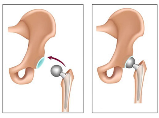
The hip joint is a ball and socket type of joint. The Total Hip Arthroplasty operation changes both the ball and the socket to artificial replacements (prosthetics/ implants).

There are a few technical consideration which make the operation performed different:

1. Material used in Artificial Joint

Cups and Stems: The cup is the replacement socket part of the new hip joint. The stem is the new ball which is attached to the thigh bone and fits into the new socket. These are usually make of titanium.

New joint surfaces - bearing surfaces: Most common these days is a Polyethelene liner - this fits inside the cup. The head surface is commonly cobalt-chrome. Ceramic and metal have also been used. The various materials have pros and cons related to their individual properties.

Ultra high molecular weight Polyethelene is a good articular surface but tends to have a high wear rate and the debris produced causes loosening of the join (aseptic loosening). Sterilisation of the material in an oxygen free environment promotes cross linking of the Polyethelene resulting in much less wear.

Cobalt-chrome is a 'supermetal' alloy. This is often used as a femoral (thigh bone) ball surface together with a Polyethelene liner to the socket.

Metal on metal implants have been used. The debris is much smaller with metal resulting in less chance of loosening but stimulating cancer is possible complication.

Ceramic material for the joint surface provides excellent wear rate but is brittle and can fracture.

The size of the ball join head is also important. A larger head provides more stability to the joint, but also results in more wear. Currently a 28 mm ball size is common.

2. Technique of fixation - cemented or un-cemented

Cemented: The cement used is called Methylmethacrylate. It is most often used in older patients. It advantage is that is creates an immediate strong and stable fixation. Cement tends to work better when compressed than when put in tension. Hence the femoral (thigh bone) component tends to do better than the acetabular cup part.

Un-cemented/ Biological: This is more popular in younger patients. Your own town grows onto to into the implant. When first implanted there needs to be another way of creating immediate fixation until the bone grows in. This can be done by using an implant a couple of millimetres bigger than the size of the bone. Alternatively, the exact size can be used and screws are used to hold it in place which the bone grows in.

3. Operative approach to the hip joint - which route to take to get to the hip joint.

The 'approach' refers to where the cut is made in the skin and which muscles and other structures are moved and cut to get to the hip joint to do the replacement. There are various pros and cons relating to the types of approach.

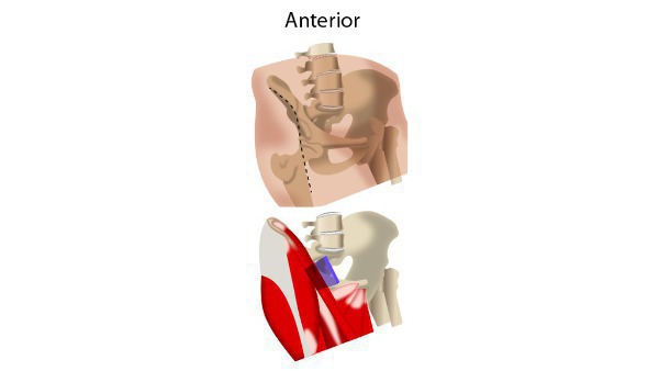
The anterior approach is when the surgeon makes a cut at the upper part of the pelvis as the patient lies on their back on the operating table. The posterior approach is when the surgeon makes a cut behind part of the upper thighbone as the patient is lying on their side (1).

The anterior approach may have earlier recovery time; a shorter incision length and reduced blood volume loss than the posterior approach (2)

Posterior approach (Moore/ Southern) to the hip joint.

This approach involves splitting the gluteus maximus muscle in the buttock and cutting a number of muscles which externally rotate the thigh bone. These are repaired at the end of the operation to prevent the hip dislocating backwards. There is a potential advantage of protection of the sciatic nerve which runs close to the hip joint at the back.


Posterior approach to the hip joint for hip arthroplasty. The blue plane shows the direction taken to get to the hip joint through the buttock area.

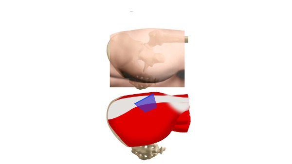
Lateral approach (Hardinge) to the hip joint.

The gluteus medius muscle is split and pushed apart to reveal the outer surface of the hip joint. This approach may result in less risk of dislocation after surgery.

Anterero-lateral approach (Watson-Jones) to the hip joint.

Here, a space between the tensor facia lata and the gluteus medius is created by pushing those two muscles apart. The abductor muscles must be detached from the femur to get to the hip joint. There is a potential risk to the femoral nerve using this approach as it needs to be pushed out of the way of the surgery.


The antero-lateral approach to the hip joint is shown. The blue plane shows the direction taken to get to the hip join from the front.

Why is it done?

Conditions that damage the hip joint to the point where you may require hip replacement surgery include: 

Osteoarthritis(OA) - Wear and tear disease of the smooth cartilage that covers the bone end at a joint helping it to move smoothly.

Rheumatoid arthritis(RA) - Autoimmune disease that causes inflation and erodes the cartilage and underlying bones resulting in deformed joints.

Osteonecrosis - Inadequate blood supply to the hip joint resulting in bone collapse and deformation.

These conditions are initially treated with medications and physiotherapy, but ultimately requires arthoplasty when hip pain: Persists, despite pain medication Worsens with walking, even with a cane or walker Interferes with your sleep Affects your ability to go up or down stairs Makes it difficult to rise from a seated position.

Will I need to do any preparation?

Your symptoms are most important in terms of decision making regarding having a hip replacement. Together with the results of clinical examination, blood test and an X-ray and possibly an MRI, an overall picture is developed regarding need for surgery.

Your bodies ability to deal with the stress of surgery will also need to be assessed. This is often done in pre-admission clinics where relevant tests and your fitness are considered.

Technique

Anaesthesia

The operation is commonly performed under general anaesthetic which the patient will be asleep during the operation. The operation can be performed using an epidural or spinal anaesthetic which only acts on the lower body.

What does it involve?

The operation can be divided into two parts, one to deal with he acetabulum and another to deal with the Femur (thigh bone).

Anterior approach

The patient is positioned so they are lying on their back so that the surgeon has access to the front of the leg. A cut is made at the top of the leg underneath one of the bony prominences on the front of the pelvis. One of the muscles (tensor fascia lata) in the leg is cut to provide access to the joint. The surrounding ligaments and fatty tissue are removed to expose the bone.

Posterior approach

The patient is positioned so they are lying on their side so that the surgeon has access to the back of the leg. A cut is made at the top of the leg.

One of the muscles (gluteus maximus) in the leg is cut to provide access to the joint. The surrounding ligaments and fatty tissue are removed to expose the bone. The socket of the joint (acetabulum) is replaced with an artificial cup.

Femoral part

The head of the femur (thighbone) is removed and an artificial head is inserted.

The femoral head (part of the thigh bone) - the 'ball' part of the joint is removed.The acetabulum (part of the hip bone) - the socket is removed also.

Acetabular part

The socket of the joint (acetabulum) is replaced with an artificial cup.


'Reducing' the joint back in place


The replaced parts of the hip joint are now 'reduced' back into place.

The wound is closed with stitches and dressed.

How long does it take? 

The operation usually takes 1-2 hours to complete.

Post procedure course (follow-up)

Surgery is followed by early mobilization, this is important to minimise complications.

Elastic compression stockings or inflatable air sleeves are worn on the legs and blood thinning medication is taken to reduce the risk of blood clotting in the legs. a major risk after Hip Arthoplasty.

How long will I stay in hospital?

People often stay in hospital for up to 3-5 days. The team will assess how the new hip is functioning and advise a personalised rehabilitation plan for the following months to help recovery. This will involve a physiotherapist and an occupational therapist.

What care will I need at home?

Some may require inpatient rehabilitation in a dedicated rehabilitation unit or nursing home. Mobilisation and physical therapy begin as soon as possible to speed up recovery and reduce risk of deep vein thrombosis (DVT). 

Enhanced recovery protocols are commonly used with targets to achieve every day. Rehabilitation exercise may involve 20 to 30 minutes two or three times daily during initially. You are allowed to weight bear as soon as tolerated is allowed.

Will I need someone to stay with me?

Depending on your level of function when you go home, you may need someone to help you with your daily activities. Your needs are professionally assessed by a physiotherapist and occupational therapist before discharge from hospital.

Will I need any special equipment when I go home?

Assist devices and walking aids (Zimmer, rolator frames or crutches) are used to help with balance and stability. Walking aids are weaned off as strength, balance, and comfort improve.

What follow up care is needed? 

6 to 12 weeks after surgery, you'll have a follow-up appointment with your surgeon to make sure your hip is healing properly. If recovery is progressing well, most people resume their normal activities by this time — even if in a limited fashion. Further recovery with improving strength will often occur for six to 12 months.

When can I start my normal activities again (e.g. driving, sports or work?)

The doctor or physiotherapist will be able to advise on when it is safe to return to normal activities which can differ from patient to patient. A rough guide is 6 weeks before returning to light activities and driving. Improvement may continue for up to a year.

Alternative treatment options

Conservative Treatment:

Patients who have severe arthritis of the hip, but function adequately, can choose to live with their condition Among these are physical therapy, walking aids, anti-inflammatory medications, cortisone injections, and joint supplements.11 Hip Resurfacing Hip resurfacing surgery is an alternative to standard hip replacements for patients with severe arthritis. In a hip resurfacing surgery, the implant is smaller, and less normal bone is removed. Hip resurfacing is gaining interest, especially in younger patients.11 Partial Hip Replacement (Hemiarthroplasty) In this procedure only ball of ball and socket joint of hip is removed good alternative in case of trauma specially in young patient but poor outcomes in hip arthritis as this procedure donot addresses the socket part of the joint.11 Hip Fusion (Arthrodesis) Hip fusion is rarely done because of the growing success of hip arthoplasty involving fusion of hip bone and femur preventing any movement at hip joint at the cost of less post operative movement rehabilitation procedures.11 Resection Arthroplasty (Gridlestone Procedure): A resection arthroplasty is a procedure where the bone around the hip joint is removed and the joint space is allowed to fill with scar tissue. This procedure is usually done in patients with a severe infection that cannot be controlled, or in patients whose physical condition is such that they have little chance of normal walking. Patients who undergo a Girdlestone resection arthroplasty will likely need some device (crutches or walker) for walking. Hip Osteotomy Hip osteotomy is a procedure that is done to realign the bones of the hip joint. The osteotomy can be performed on the thigh bone (femur), the pelvis, or both. Osteotomies are usually performed on patients with an underlying problem that has led to early arthritis of the hip joint. For example, developmental conditions such as hip dysplasia can lead to early hip arthritis. Hip dysplasia occurs in infants and leads to misaligned bones around the hip. The osteotomy helps to realign the bones and relieve the symptoms of early arthritis. These procedures must be done in carefully selected patients.


More from this author:

prof. Usman Jafferprof. Usman Jaffer

Knee Replacement

prof. Usman Jafferprof. Usman Jaffer

Ankle Arthroscopy

Indication: this procedure is used for diagnosis as well as theraputic purpose thus indication is divided into Diagnostic indication: 1) Pain, swelling, stiffness, instability of the ankel joint that remain unexplained. 2) locking and popping symptoms of ankle joint Therapeutic Indication: 1) articular or soft tissue injury. 2) soft tissue or bone impingement and instability. 3) Inflammation of lining membranes of the joint(Synovitis). 4) inflammation of tendons (tendinitis) 5) Joint infection (septic arthritis) 6) Surgical immobilization of joint (arthrodesis) 7) Adhesion inside joints (intra articular bands) Contraindication: Absolute contraindication: 1) Active local soft tissue infection. 2) Severe degenerative (Wear & tear) joint disease. 3) Poor vascular supply the leg. Relative Contraindication: 1) Moderate degenerative (wear and tear) joint disease. 2) Severe Edema (swelling). Procedure: it is usually performe when conservative measurement fails to treat the underlying condition.These includes use of pain medication like (NSAIDS), Ankle braces, heel lift or wedge Patient preparation involve history examination and blood test including CBC(complet blood count) with ESR(erythrocyte sedimentation rate) , C-reactive protein (CRP), coagulation profile , in case of infection joint aspiration is done for culture and sensitivity, radiological investigation like X-ray, Ct-scan and MRI. After proper analgesia and anesthesia either local or Regional, a blood less field is achieved by the use of the tourniquet just above the ankle or by using high inflow out flow system that also improve visualization and irrigate the debris material.Small incisions are then made for portals, which are small tubes which are placed in different area around ankle for the instruments and camera to be placed in. after that the surgeon the perform the procedure like removal of inflamed synovial membranes and debris after ankle joint soft tissue or bone injury. at the end the portals and instruments are removed and the small incisions are stitched closed and bandaged. After procedure depending upon the patient condition and surgeons choice some are allowed to bear weight with crutches while others may be placed in an immobilizer for as long as six weeks.In case of extensive surgery or remodeling ankle is put in cast to prevent early mobilization and promote healing and if arthroscopy is performed only for diagnostic purpose simple splint is enough.

prof. Usman Jafferprof. Usman Jaffer

Anterior cruciate ligament repair

Reconstruction surgeries are of two type arthroscopic and open reconstruction depending on orthopedic surgeon’s choice and extent of injury. Time of surgery is debatable topic. Surgery immediately after injury has been associated with increased fibrous tissue leading to loss of motion (arthofibrosis) after surgery (1). According to some surgeon surgery should be delayed until swelling goes down, gain range of motion in knee and can strongly contract (flex) front of thigh muscle (quadriceps)(1). In adults age is not a factor in surgery though overall condition of body may be. People with medical conditions may consider non surgical treatment option because surgery carries greater risk. These include use of painkillers and anti inflammatory medications and bracing for 1 to 2 weeks with ice, elevation and programmed physiotherapy. A new non surgical treatment option is Platelet rich plasma injection, in this procedure blood of the patient is taken platelets are separated by centrifuge and injected I the injured tissue to augment healing. It is useful in both acute and chronic soft tissues injuries. Choice completely depends upon patient choice and level of activity patient having after surgery (2). Many orthopedic surgeons use arthroscopic surgery rather than open surgery for ACL injuries because: 1. It is easy to see and work on the knee structures. 2. It uses smaller incisions than open surgery. 3. It can be done at the same time as diagnostic arthroscopy (using arthroscopy to find out about the injury or damage to the knee). 4. It may have fewer risks than open surgery (3). During arthroscopic ACL reconstruction, the surgeon makes several small incisions—usually two or three—around the knee. Sterile saline (salt) solution is pumped into the knee through one incision to expand it and to wash blood from the area. This allows the doctor to see the knee structures more clearly. The surgeon inserts an arthroscope into one of the other incisions. A camera at the end of the arthroscope transmits pictures from inside the knee to a TV monitor in the operating room. Surgical drills are inserted through other small incisions. The surgeon drills small holes into the upper and lower leg bones where these bones come close together at the knee joint. The holes form tunnels through which the graft will be anchored. If you are using your own tissue, the surgeon will make another incision in the knee and take the graft (replacement tissue). The graft is pulled through the tunnels that were drilled in the upper and lower leg bones. The surgeon secures the graft with hardware such as screws or staples and will close the incisions with stitches or tape. The knee is bandaged, and you are taken to the recovery room for 2 to 3 hours.3 Physical rehabilitation after ACL surgery may take several months to a year. The length of time until you can return to normal activities or sports is different for every person. It takes most people at least 6 months to return to activity after surgery (4). ACL reconstructive surgery remains the gold standard for repairing this common knee injury. The AAOS reports that about 82 to 90 percent of ACL reconstruction surgeries yield excellent results and full knee stability. ACL reconstruction surgery is generally safe. Complications that may arise from surgery or during rehabilitation (rehab) and recovery include following (6, 7). 0.75% (104) of the consecutive patient cohort had a wound complication recorded (5). • Problems related to the surgery itself. These are uncommon but may include:  Numbness in the surgical scar area.  Infection in the surgical incisions. 0.25% (35) underwent a further procedure to wash out the infected knee joint (5).  Damage to structures, nerves, or blood vessels around and in the knee.  Blood clots in the leg. DVT and PTE rates were 0.30% (42) and 0.18% (25) respectively with on 90 days(5)  The usual risks of anesthesia.  There were no in-hospital deaths(5) • Problems with the graft tendon (loosening, stretching, re-injury, or scar tissue). The screws that attach the graft to the leg bones may cause problems and require removal. • Limited range of motion, usually at the extremes. For example, you may not be able to completely straighten or bend your leg as far as the other leg. This is uncommon, and sometimes another surgery or manipulation under anesthesia can help. Rehab attempts to restore a range of motion between 0 degrees (straight) and 130 degrees (bent or flexion). It's important to be able to get your knee straight so you can walk normally. • Grating of the kneecap (crepitus) as it moves against the lower end of the thigh bone (femur), this may develop in people who did not have it before surgery. This may be painful and may limit your athletic performance. In rare cases, the kneecap may be fractured while the graft is being taken during surgery or from a fall onto the knee soon after surgery. • Pain, when kneeling, at the site where the tendon graft was taken from the patellar tendon or at the site on the lower leg bone (tibia) where a hamstring or patellar tendon graft is attached. • Repeat injury to the graft (just like the original ligament). Repeat surgery is more complicated and less successful than the first surgery. 1.36% (190) were readmitted to an orthopedic ward within 30 days (5)

prof. Usman Jafferprof. Usman Jaffer

Femoral- Above the knee Popliteal Bypass Graft

prof. Usman Jafferprof. Usman Jaffer

Femoral-Below knee Popliteal Artery Bypass Graft

prof. Usman Jafferprof. Usman Jaffer

Anal Fistula

Treatment of anal fistula: A fistula without symptoms found on regular check up usually requires no therapy . [3,4] In case of anorectal abscess surgery for fistula repair should not be performed (unless the fistula is superficial and the tract is obvious). In the acute phase, simple incision and drainage of the abscess are sufficient. [5] Only 7-40% of patients will develop a fistula. Recurrent anal sepsis and fistula formation are twofold higher after an abscess in patients younger than 40 years and are almost threefold higher in non diabetics. Preoperative considerations include the following: • Rectal irrigation with enemas should be performed on the morning of the operation • Anesthesia can be general, local with intravenous sedation, or a regional block • Administer preoperative antibiotics Intraoperative considerations include the following: • Examine the patient under anesthesia to confirm the extent of the fistula • Identifying the internal opening (inside the anus) to prevent recurrence is imperative • A local anesthetic block at the end of the procedure provides postoperative analgesia Fistulotomy: The laying-open technique (fistulotomy) is useful for 85-95% of primary fistulas (ie, submucosal, intersphincteric, and low transsphincteric). [6, 7, 8, 9] The most common type of surgery for anal fistulas is a fistulotomy. This involves cutting along the whole length of the fistula to open it up so it heals as a flat scar.A fistulotomy is the most effective treatment for many anal fistulas, although it's usually only suitable for fistulas that don't pass through much of the sphincter muscles, as the risk of incontinence is lowest in these cases. If your surgeon has to cut a small portion of anal sphincter muscle during the procedure, they will make every attempt to reduce the risk of incontinence.In cases where the risk of incontinence is considered too high, one of the procedures below may be recommended instead. Seton techniques: If your fistula passes through a significant portion of anal sphincter muscle, your surgeon may initially recommend inserting a seton. A seton is a piece of surgical thread that is left in the fistula for several weeks to keep it open. This allows it to drain and helps it heal, while avoiding the need to cut the sphincter muscles. Loose setons allow fistulas to drain, but don't cure them. To cure a fistula, tighter setons may be used to cut through the fistula slowly. This may require several procedures that your surgeon can discuss with you. Seton Placement: A seton can be placed alone, combined with fistulotomy, or in a staged fashion. This technique is useful in patients with the following conditions [10, 11, 12] : • Complex fistulas (ie, high transsphincteric, suprasphincteric, extrasphincteric) or multiple fistulas • Recurrent fistulas after previous fistulotomy • Anterior fistulas in female patients • Poor preoperative sphincter pressures • Patients with Crohn disease or patients who are immunosuppressed Single-stage seton (cutting): Pass the seton through the fistula tract around the deep external sphincter after opening the skin, subcutaneous tissue, internal sphincter muscle, and subcutaneous external sphincter muscle. The seton is tightened down and secured with a separate silk tie. With time, fibrosis occurs above the seton as it gradually cuts through the sphincter muscles and essentially exteriorizes the tract. The seton is tightened on subsequent office visits until it is pulled through over 6-8 weeks. A cutting seton can also be used without associated fistulotomy. Recurrence and incontinence are important factors to consider when this technique is employed. The success rates for cutting setons range from 82-100%; however, long-term incontinence rates can exceed 30%. [13,14, 15] Two-stage seton (draining/fibrosing): Pass the seton around the deep portion of the external sphincter after opening the skin, subcutaneous tissue, internal sphincter muscle, and subcutaneous external sphincter muscle. Unlike the cutting seton, the seton is left loose to drain the intersphincteric space and to promote fibrosis in the deep sphincter muscle. Once the superficial wound is healed completely (2-3 months later), the seton-bound sphincter muscle is divided. Two studies (74 patients combined) supported the two-stage approach with a 0-nylon seton. Once wound healing is complete, the seton is removed without division of the remaining encircled deep external sphincter muscle. The researchers reported eradication of the fistula tract in 60-78% of cases. Mucosal Advancement Flap: A mucosal advancement flap is reserved for use in patients with chronic high fistula but is indicated for the same disease process as seton use. [1,16, 17] Advantages include a one-stage procedure with no additional sphincter damage. A disadvantage is poor success in patients with Crohn disease or acute infection. This procedure involves total fistulectomy, with removal of the primary and secondary tracts and completes excision of the internal opening. A rectal mucomuscular flap with a wide proximal base (two times the apex width) is raised. The internal muscle defect is closed with an absorbable suture, and the flap is sewn down over the internal opening so that its suture line does not overlap the muscular repair. Plugs and Adhesives: Advances in biotechnology have led to the development of many new tissue adhesives and biomaterials formed as fistula plugs. By their less-invasive nature, these therapies lead to decreased postoperative morbidity and risk of incontinence, but long-term data are lacking for eradication of disease, especially in complex fistulas, which carry high recurrence rates. [18, 19, 17] Reported series exist of fibrin glue treatment of fistula-in-ano, with 1-year follow-up showing recurrence rates approaching 40-80%. [20, 21,22] The Surgisis fistula plug has also had mixed long-term results in direct clinical trials. [23, 24, 25] Early success rates have been reported for newer materials, such as acellular dermal matrix and the bioabsorbable Gore Bio-A fistula plug, in low fistulas and good animal model data. [26] Assessment of long-term success rates with plug techniques for complex disease will be based on further data from randomized trials. In a randomized, controlled study designed to evaluate the efficacy and safety of the anal fistula plug in patients with fistulizing anoperineal Crohn disease, Senéjoux et al did not find the plug to be superior to seton removal for achieving fistual closure, regardless of whether the fistula was simple or complex. [27] A combined sphincter-sparing repair that includes both an anal fistula plug and a rectal advancement flap has been proposed for the treatment of transsphincteric fistula-in-ano. [28] LIFT Procedure: Ligation of the intersphincteric fistula tract (LIFT) is a sphincter-sparing procedure for complex transsphincteric fistulas first described in 2007. It is performed by accessing the intersphincteric plane with the goal of performing a secure closure of the internal opening and by removing the infected cryptoglandular tissue. [29] The intersphincteric tract is identified and isolated by performing meticulous dissection through the intersphincteric plane after making a small incision overlying the probe connecting the external and internal openings. Once isolated, the intersphincteric tract is hooked with a small right-angle clamp, and the tract is ligated close to the internal sphincter and then divided distal to the point of ligation. Hydrogen peroxide is injected through the external opening to confirm the division of the correct tract. The external opening and the remnant fistulous tract are curetted to the level of the proximity of the external sphincter complex. Finally, the intersphincteric incision is loosely reapproximated with an absorbable suture. The curettaged wound is left opened for dressing.[29,30,31] Because of its relative novelty, LIFT has not been extensively researched. In a randomized trial of 39 patients with complex fistula-in-ano who had failed previous procedures and were treated with LIFT technique, success rates were comparable to those seen with the anorectal advancement flap technique. [32] The probability of recurrence at 19 months was 8% for the LIFT technique versus 7% for the anorectal advancement flap. Time to return to work was shorter in the LIFT group (1 vs 2 wk), but there was no difference in incontinence scores. [32] Further randomized surgical trials are needed to determine whether this technique is a viable—or, possibly, a better—alternative to the other previously mentioned procedures for the treatment of fistula-in-ano. Diversion: In rare cases, the creation of a diverting stoma may be indicated to facilitate the treatment of a complex persistent fistula-in-ano. The most common indications include, but are not limited to, patients with perineal necrotizing fasciitis, severe anorectal Crohn disease, reoperative rectovaginal fistulas, and radiation-induced fistulas. Fecal diversion alone is effective in these select patients to control sepsis and symptoms; however, long-term success rates after reanastomosis are low because of recurrence from the underlying disease. Thus, this approach should be avoided unless the underlying fistula-in-ano disease process is repaired or has healed completely, which is unlikely. Postoperative Care: After the operation, most patients can be treated in an ambulatory setting with discharge instructions and close follow-up care. Sitz baths, analgesics, and stool-bulking agents (eg, bran and psyllium products) are used in follow-up care. Complications: Early postoperative complications may include the following: • Urinary retention • Bleeding • Fecal impaction • Thrombosed hemorrhoids Delayed postoperative complications may include the following: • Recurrence • Incontinence (stool) • Anal stenosis - The healing process causes fibrosis of the anal canal; bulking agents for stool help to prevent narrowing • Delayed wound healing - Complete healing occurs by 12 weeks unless an underlying disease process is present (ie, recurrence, Crohn disease) Postoperative rates of recurrence and incontinence vary according to the procedure performed, as follows: • Standard fistulotomy - The reported rate of recurrence is 0-18%, and the rate of any stool incontinence is 3-7% • Seton use - The reported rate of recurrence is 0-17%, and the rate of any incontinence of stool is 0-17% • Mucosal advancement flap - The reported rate of recurrence is 1-17%, and the rate of any incontinence of stool is 6-8% [16] Long-Term Monitoring: Frequent office visits within the first few weeks help to ensure proper healing and wound care. It is important to ensure that the internal wound does not close prematurely, causing a recurrent fistula. Digital examination findings can help distinguish early fibrosis. Wound healing usually occurs within 6 weeks.