Health Shared Logo whiteHealth Shared Logo dark

ERCP (Endoscopic retrograde cholangiopancreatography)

prof. Usman Jafferprof. Usman Jaffer

Introduction

What is it?

Endoscopic retrograde cholangiopancreatography or ‘ERCP’ is a procedure that is used to look at the drainage channels (bile ducts) arising from your liver and pancreas. It is performed by a specially trained doctor called an endoscopist to diagnose and treat conditions of the biliary tract. Treatment of conditions using ERCP depends on the problem. For example, gallstones can be removed using a small cut to the duct to allow passage of stones, or stents can be placed in bile ducts where there is a narrowing.

What is the biliary tract?  

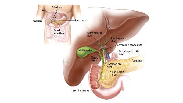
The biliary tract contains the organs and channels that produce and store bile. The main role of the biliary tract is to allow the flow of bile from the liver into the first part of the small bowel (the duodenum) where it helps in the digestion of food. Some bile is stored in the gallbladder for later use. The biliary tract starts with many small branches in the liver which join together to form bigger branches eventually all leading to a channel called the common bile duct. For this reason, it is sometimes known as the ‘biliary tree’. The common bile duct allows bile to flow into the small bowel through a small hole called the papilla. This is the point where the pancreatic duct also allows pancreatic juice to enter the small bowel.


Top leg: Location of the biliary tract (under the Liver). Right: The Liver, Gall bladder and bile duct - emptying into t he small intestine. The pancreases and pancreatic duct - also. emptying into the. intestine.

What is Bile? 

The liver produces around 600ml of bile a day. Bile is a yellowish green liquid which contains substances needed for digestion of fats in particular. Bile flows from the liver into the digestive tract through a system of channels called bile ducts. Bile is responsible for the brown colouration of our faeces. In some conditions if there is a blockage of bile reaching the intestine, faeces can become pale/whitish as not enough bile is reaching the gut and instead overflows into the blood steam resulting in yellowing of the skin (jaundice)

Why is it done?

An ERCP is a procedure that can be used to diagnose and treat certain conditions of the biliary system. Conditions of the biliary system can result in a number of symptoms including jaundice (yellowing of the skin), weight loss, abnormal bowels, abdominal pains, and infections. 

An ERCP in most cases is performed when evidence of abnormal blood tests or scans of the abdomen (ultrasound or CT) point to cause. The most common reason for an ERCP is for the treatment of gallstones causing a blockage of the bile ducts. Often this is the situation when pre-operative tests are being done for a patient about to have a laparoscopic cholecystectomy.

Other indications for ERCP (from the British Society of Gastroenterology guidelines): 1. Bile duct stones 2. Jaundice due to a stricture (narrowing) of the lower bile ducts in patients not suitable for an operation 3. Hilar biliary stricture (narrowing of the bile ducts entering the liver) 4. Bile leak or jaundice following a cholecystectomy (gall bladder surgery) 5. Pancreatic duct stricture (narrowing) causing symptoms 6. For biopsies 7. Emergency treatment of cholangitis (infection of the bile ducts causing sepsis

Will I need to do any preparation?

Preassessment Clinic You may be seen by a nurse or doctor in a pre assessment clinic a few weeks prior to the ERCP. Your full medical history will be obtained and your suitability for the procedure will be determined following this. Any medications that need to be stopped or changed will also be addressed. Patients with the following conditions should ensure they let the doctor know prior to the procedure • Diabetes • Haemophilia or other blood disorders • Pacemaker or other implant • Blood thinning medications • Heart or lung diseases Pregnancy 

Although ERCP can be performed safely during pregnancy, this is usually reserved for urgent indications. Most non urgent cases are postponed until after child birth. Medications Some medications may need to be stopped prior to having an ERCP. Blood thinning medications (e.g Aspirin, Clopidogrel, Warfarin, Enoxaparin, Rivaroxiban, Apixiban etc..) may need to be held to reduce the risk of bleeding during and after the procedure. You should discuss this with the doctor well in advance of the ERCP to allow time to plan. Sometimes the doctor will need to take advice from other specialists when holding certain mediations. Certain diabetic medications may also need to be stopped or adjusted on the morning of the procedure as you will not be eating for 6-8 hours. On the day Nil by mouth You will be asked to remain ‘Nil by mouth’ for six to eight hours before the procedure. This means you should not eat or drink during this time. This is to allow the doctor to visualise the structures clearly (without food being present in the gut) and also to reduce the risk of vomiting.

Technique

Anaesthesia

You will be given a local anaesthetic throat spray to help numb your throat. This also decreases the gag reflex when the endoscope is passed into your food pipe. In most cases conscious sedation is used, however in some units and in complex cases, a general anaesthetic is used.

Sedation 

Most ERCPs are performed under ‘sedation’. An intravenous cannula (small tube to allow administration of medications) will be inserted into one of the veins in your hand or arm. Most endoscopy units use a sedative medication to cause relaxation in addition to medication to relieve pain. This is called ‘conscious sedation’ as you will not be asleep.

General Anaesthetic

Some units and in ore complex cases you may require a general anaesthetic – where an anaesthetist will use medication to put you asleep.

What does it involve?

Before the Procedure: 1. Arrival to the Endoscopy Unit. If you are an inpatient you will usually be escorted by a nurse to the endoscopy unit. This is a designated part of the hospital containing rooms that are used for endoscopic procedures. If you are an out-patient you will be asked to report to the unit at a specific time. You may have to wait in a waiting area until you are seen by an endoscopy nurse who will go through a few identity checks and medical history with you. You will be given a hospital gown to change into. 

2. Consent Form Before the ERCP you will be seen by the endoscopist who will also go through your medical history, blood results and any recent scans that you may have had. The endoscopist will explain the procedure in full, discussing the benefits and risks of ERCP. You will be asked to sign a consent form.

3. Endoscopy room. ERCP is performed in an endoscopy room containing x ray equipment. This is because as describe earlier, x rays are used to examine the bile ducts following injection of a special dye into them. The room will also contain monitors which are connected to the camera on the endoscope which is essential for the doctor to visualise the digestive tract.

4. Monitoring You will be attached to some monitors before the procedure, to measure your blood pressure, heart rate, heart rhythm and oxygen levels.

The Duodenoscope 

The ERCP endoscope is called a duodenoscope. It is an advanced flexible tube which is approximately the same width of an index finger. It contains a small camera and a light allowing the doctor to see the inside of the digestive tract on a monitor. The endoscope contains channels that allow insertion of a variety of instruments – to take biopsies, withdraw fluid, introduce air, introduce cutting devices and stents.

The procedure:

1. You will be asked to lie on a special bed on your left side or belly.


Usual position for ERCP with patient lying on one side. The room contains an endoscopy machine and an X ray machine.

2. A mouth guard is inserted between the teeth – this prevents damage to the teeth and the endoscope.


Mouth guard to protect the teeth from the endoscope as it is passed in the mouth and food pipe.


3. The endoscope will be inserted into the mouth and you will then be asked to swallow it. Most people do not have difficulty with this due to the sedating mediations given. One the endoscope has been inserted, air is gently introduced to inflate the oesophagus, stomach and intestine so that it can be passed through with ease. 4. Once the papilla has been reached (the opening of the bile ducts into the intestine) a cannula (small plastic tube) is inserted through this opening and a special dye is injected.

5. X rays are taken after the dye has been injected which allow examination of the bile ducts and pancreatic duct.


X ray picture of an ERCP being done. The large snake like object is the endoscope (duodenoscope). Bottom right: X ray dye is being injected into the ampulla which is outlining the biliary tract.

How long does it take? 

6. If any intervention or treatment is required, sometimes a sphinterotomy is required. A spincterotomy involves cutting of the muscle at the mouth of the papilla. This allows for the hole to be widened up an allow instruments to be passed into the ducts.

How long does it take? 

The procedure takes between 30 mins to over 3 hours, depending on the procedures or complexity of the case. Longer treatment times are usually if procedures like stone extraction or stent placement are done.

Post procedure course (follow-up)

When can I eat and Drink? 

Usually you may eat and drink once you are fully awake, however in some cases you may be required to continue to fast for a further period of time. The doctor performing the procedure will write a set of notes detailing your post procedure care which would have been given to the nurses and doctors looking after you.

Do I need someone to take me home?

If you are going home you will require someone to escort you as the sedation takes time to wear off you will not be able to drive.

Should I take my usual medications after the procedure? 

You should continue to take your usual medications unless instructed by your doctor. If any medications have been stopped – the doctor will inform you when it is safe to restart these.

Can I drink Alcohol? 

You should not drink alcohol for at least 24 hours after the procedure as the sedation can take time to wear off. Discuss this with your doctor.

How long will I stay in hospital?

Following the procedure you will be monitored in the recovery area by nurse who will assess your blood pressure, oxygen levels, temperature and heart rate regularly. You will usually remain in the unit until you are fully awake which takes about one hour. Following this you will be observed for a further 6 hours following the procedure

What care will I need at home?

No particular care required.

Will I need someone to stay with me?

It is advisable to have somebody with you for at least 24 hours until the sedation has worn off.

Will I need any special equipment when I go home?

No routine special equipment us needed unless otherwise you are told otherwise.

What follow up care is needed? 


When can I start my normal activities again (e.g. driving, sports or work?)

Sedation can sometimes last longer than you think. During the first 24 hours after the procedure you should not drive, ride a bike, operate machinery or do anything that requires a skill. You should also not drink alcohol, take sleeping tablets or make important decisions.


More from this author:

prof. Usman Jafferprof. Usman Jaffer

Cholecystectomy

A cholecystectomy is mostly performed to treat gallstones which are small stone formed for no reasons in the gallbladder consisting of cholesterol or bile salt. Risk factors include 4F i.e Female, Fertile Forty and Family (history). These stone may cause blockage of bile duct in turn causing and irritation of gallbladder resulting in sharp pain in the abdomen in the right upper quadrant associated with vomiting, indigestion and sometimes fever. Cholecystectomy is usually performed through a minimal invasive procedure known as laproscopic cholecystectomy. Open cholecystectomy is the choice of the surgeon either before or during the laproscopic cholecytectomy strictly based on patient safety. Cholecystectomy is primarily indicated in following conditions; • Stone in gall bladder (cholelithiasis) • Stone in the bile duct(choledocolithiasis) • Gallbladder calcification (porcelain gallbladder) • Inflammation of gall bladder (cholecystitis) • Inflammation of pancrease due to stone blockage (biliary pancreatitis) • Lack of movement of bilary channels (bilary dyskinesia) • Gallbladder cancer Indications for Consideration of Prophylactic Cholecystectomy • Risk factors for carcinoma.1 • Anomalous pancreatic–biliary ductal junctions.[1] • Choledochal cysts.[1] • Gallbladder adenomas.1 • Native North or South American background.2 • Porcelain gallbladder.1 • Solitary gallbladder polyp larger than 1 cm.2 Other indications • Choledocholithiasis.2 • Gallstones larger than 3 cm.2 • Patient lives in remote location from health care facility.2 • Sickle cell disease/spherocytosis.1 • Transplant or immunosuppressant therapy.2 • Young age.3 How to perform the procedure: Under general anesthesia, so the patient is asleep throughout the procedure. Using a cannula (a narrow tube-like instrument), the surgeon enters the abdomen in the area of the belly-button.A laparoscope (a tiny telescope) connected to a special camera is inserted through the cannula, giving the surgeon a magnified view of the patient’s internal organs on a television screen. Other cannulas are inserted which allow your surgeon to delicately separate the gallbladder from its attachments and then remove it through one of the openings. An X-ray called a cholangiogram may be performed during the operation to identify stones which may be lodged in the bile channels or to verify the anatomy of structures that have been identified. If the surgeon finds one or more stones in the common bile duct, he/she may remove them with a special scope, may choose to have them removed later through an endoscope placed through the mouth into the stomach using a procedure called ERCP or may convert to an open operation in order to remove all the stones during the operation.After the surgeon removes the gallbladder, the small incisions are closed with absorbable stitches or with surgical tape or glue.

prof. Usman Jafferprof. Usman Jaffer

Anal Fistula

Treatment of anal fistula: A fistula without symptoms found on regular check up usually requires no therapy . [3,4] In case of anorectal abscess surgery for fistula repair should not be performed (unless the fistula is superficial and the tract is obvious). In the acute phase, simple incision and drainage of the abscess are sufficient. [5] Only 7-40% of patients will develop a fistula. Recurrent anal sepsis and fistula formation are twofold higher after an abscess in patients younger than 40 years and are almost threefold higher in non diabetics. Preoperative considerations include the following: • Rectal irrigation with enemas should be performed on the morning of the operation • Anesthesia can be general, local with intravenous sedation, or a regional block • Administer preoperative antibiotics Intraoperative considerations include the following: • Examine the patient under anesthesia to confirm the extent of the fistula • Identifying the internal opening (inside the anus) to prevent recurrence is imperative • A local anesthetic block at the end of the procedure provides postoperative analgesia Fistulotomy: The laying-open technique (fistulotomy) is useful for 85-95% of primary fistulas (ie, submucosal, intersphincteric, and low transsphincteric). [6, 7, 8, 9] The most common type of surgery for anal fistulas is a fistulotomy. This involves cutting along the whole length of the fistula to open it up so it heals as a flat scar.A fistulotomy is the most effective treatment for many anal fistulas, although it's usually only suitable for fistulas that don't pass through much of the sphincter muscles, as the risk of incontinence is lowest in these cases. If your surgeon has to cut a small portion of anal sphincter muscle during the procedure, they will make every attempt to reduce the risk of incontinence.In cases where the risk of incontinence is considered too high, one of the procedures below may be recommended instead. Seton techniques: If your fistula passes through a significant portion of anal sphincter muscle, your surgeon may initially recommend inserting a seton. A seton is a piece of surgical thread that is left in the fistula for several weeks to keep it open. This allows it to drain and helps it heal, while avoiding the need to cut the sphincter muscles. Loose setons allow fistulas to drain, but don't cure them. To cure a fistula, tighter setons may be used to cut through the fistula slowly. This may require several procedures that your surgeon can discuss with you. Seton Placement: A seton can be placed alone, combined with fistulotomy, or in a staged fashion. This technique is useful in patients with the following conditions [10, 11, 12] : • Complex fistulas (ie, high transsphincteric, suprasphincteric, extrasphincteric) or multiple fistulas • Recurrent fistulas after previous fistulotomy • Anterior fistulas in female patients • Poor preoperative sphincter pressures • Patients with Crohn disease or patients who are immunosuppressed Single-stage seton (cutting): Pass the seton through the fistula tract around the deep external sphincter after opening the skin, subcutaneous tissue, internal sphincter muscle, and subcutaneous external sphincter muscle. The seton is tightened down and secured with a separate silk tie. With time, fibrosis occurs above the seton as it gradually cuts through the sphincter muscles and essentially exteriorizes the tract. The seton is tightened on subsequent office visits until it is pulled through over 6-8 weeks. A cutting seton can also be used without associated fistulotomy. Recurrence and incontinence are important factors to consider when this technique is employed. The success rates for cutting setons range from 82-100%; however, long-term incontinence rates can exceed 30%. [13,14, 15] Two-stage seton (draining/fibrosing): Pass the seton around the deep portion of the external sphincter after opening the skin, subcutaneous tissue, internal sphincter muscle, and subcutaneous external sphincter muscle. Unlike the cutting seton, the seton is left loose to drain the intersphincteric space and to promote fibrosis in the deep sphincter muscle. Once the superficial wound is healed completely (2-3 months later), the seton-bound sphincter muscle is divided. Two studies (74 patients combined) supported the two-stage approach with a 0-nylon seton. Once wound healing is complete, the seton is removed without division of the remaining encircled deep external sphincter muscle. The researchers reported eradication of the fistula tract in 60-78% of cases. Mucosal Advancement Flap: A mucosal advancement flap is reserved for use in patients with chronic high fistula but is indicated for the same disease process as seton use. [1,16, 17] Advantages include a one-stage procedure with no additional sphincter damage. A disadvantage is poor success in patients with Crohn disease or acute infection. This procedure involves total fistulectomy, with removal of the primary and secondary tracts and completes excision of the internal opening. A rectal mucomuscular flap with a wide proximal base (two times the apex width) is raised. The internal muscle defect is closed with an absorbable suture, and the flap is sewn down over the internal opening so that its suture line does not overlap the muscular repair. Plugs and Adhesives: Advances in biotechnology have led to the development of many new tissue adhesives and biomaterials formed as fistula plugs. By their less-invasive nature, these therapies lead to decreased postoperative morbidity and risk of incontinence, but long-term data are lacking for eradication of disease, especially in complex fistulas, which carry high recurrence rates. [18, 19, 17] Reported series exist of fibrin glue treatment of fistula-in-ano, with 1-year follow-up showing recurrence rates approaching 40-80%. [20, 21,22] The Surgisis fistula plug has also had mixed long-term results in direct clinical trials. [23, 24, 25] Early success rates have been reported for newer materials, such as acellular dermal matrix and the bioabsorbable Gore Bio-A fistula plug, in low fistulas and good animal model data. [26] Assessment of long-term success rates with plug techniques for complex disease will be based on further data from randomized trials. In a randomized, controlled study designed to evaluate the efficacy and safety of the anal fistula plug in patients with fistulizing anoperineal Crohn disease, Senéjoux et al did not find the plug to be superior to seton removal for achieving fistual closure, regardless of whether the fistula was simple or complex. [27] A combined sphincter-sparing repair that includes both an anal fistula plug and a rectal advancement flap has been proposed for the treatment of transsphincteric fistula-in-ano. [28] LIFT Procedure: Ligation of the intersphincteric fistula tract (LIFT) is a sphincter-sparing procedure for complex transsphincteric fistulas first described in 2007. It is performed by accessing the intersphincteric plane with the goal of performing a secure closure of the internal opening and by removing the infected cryptoglandular tissue. [29] The intersphincteric tract is identified and isolated by performing meticulous dissection through the intersphincteric plane after making a small incision overlying the probe connecting the external and internal openings. Once isolated, the intersphincteric tract is hooked with a small right-angle clamp, and the tract is ligated close to the internal sphincter and then divided distal to the point of ligation. Hydrogen peroxide is injected through the external opening to confirm the division of the correct tract. The external opening and the remnant fistulous tract are curetted to the level of the proximity of the external sphincter complex. Finally, the intersphincteric incision is loosely reapproximated with an absorbable suture. The curettaged wound is left opened for dressing.[29,30,31] Because of its relative novelty, LIFT has not been extensively researched. In a randomized trial of 39 patients with complex fistula-in-ano who had failed previous procedures and were treated with LIFT technique, success rates were comparable to those seen with the anorectal advancement flap technique. [32] The probability of recurrence at 19 months was 8% for the LIFT technique versus 7% for the anorectal advancement flap. Time to return to work was shorter in the LIFT group (1 vs 2 wk), but there was no difference in incontinence scores. [32] Further randomized surgical trials are needed to determine whether this technique is a viable—or, possibly, a better—alternative to the other previously mentioned procedures for the treatment of fistula-in-ano. Diversion: In rare cases, the creation of a diverting stoma may be indicated to facilitate the treatment of a complex persistent fistula-in-ano. The most common indications include, but are not limited to, patients with perineal necrotizing fasciitis, severe anorectal Crohn disease, reoperative rectovaginal fistulas, and radiation-induced fistulas. Fecal diversion alone is effective in these select patients to control sepsis and symptoms; however, long-term success rates after reanastomosis are low because of recurrence from the underlying disease. Thus, this approach should be avoided unless the underlying fistula-in-ano disease process is repaired or has healed completely, which is unlikely. Postoperative Care: After the operation, most patients can be treated in an ambulatory setting with discharge instructions and close follow-up care. Sitz baths, analgesics, and stool-bulking agents (eg, bran and psyllium products) are used in follow-up care. Complications: Early postoperative complications may include the following: • Urinary retention • Bleeding • Fecal impaction • Thrombosed hemorrhoids Delayed postoperative complications may include the following: • Recurrence • Incontinence (stool) • Anal stenosis - The healing process causes fibrosis of the anal canal; bulking agents for stool help to prevent narrowing • Delayed wound healing - Complete healing occurs by 12 weeks unless an underlying disease process is present (ie, recurrence, Crohn disease) Postoperative rates of recurrence and incontinence vary according to the procedure performed, as follows: • Standard fistulotomy - The reported rate of recurrence is 0-18%, and the rate of any stool incontinence is 3-7% • Seton use - The reported rate of recurrence is 0-17%, and the rate of any incontinence of stool is 0-17% • Mucosal advancement flap - The reported rate of recurrence is 1-17%, and the rate of any incontinence of stool is 6-8% [16] Long-Term Monitoring: Frequent office visits within the first few weeks help to ensure proper healing and wound care. It is important to ensure that the internal wound does not close prematurely, causing a recurrent fistula. Digital examination findings can help distinguish early fibrosis. Wound healing usually occurs within 6 weeks.

prof. Usman Jafferprof. Usman Jaffer

Splenectomy

prof. Usman Jafferprof. Usman Jaffer

Laparoscopic Cholecystectomy

prof. Usman Jafferprof. Usman Jaffer

Flexible Sigmoidoscopy

prof. Usman Jafferprof. Usman Jaffer

Anal fissure

An anal fissure is a tear or open sore (ulcer) that develops in the lining of the large intestine, near the anus. Anal fissures are quite common, with around 1 in every 10 people affected at some point in their life. They affect both sexes equally and people of all ages can get them. But children and young adults between 10 and 30 years of age are more likely to get anal fissures.[5] Anal fissure symptoms The most common symptoms of anal fissures are: • Sharp excruciating burning pain on defecation. • Minute bleeding resulting in bright red spot on feces and toilet paper Most people delay treatment because of embracement resulting in complications but a timely visit to a GP results in early recovery on conservative management. Diagnosing anal fissures Anal fissure are solely diagnose on clinical history and examination. History includes inquiry about sign and symptoms as well as bowl habits and inquiry about general health. Examination is done slightly separating the buttocks and ulcer can be visualized around anal rim under proper illumination. Digital rectal examination procedure in which your doctor will insert a gloved finger in the anus to examine in inside of the anal canal and rectum. However it is avoided because of excruciating pain that patient experience an deserve only of complicated cases and preferably done under local anesthesia to reduce pain during examination What causes anal fissures? Anal fissures are caused by damage to the anal canal lining resulting in tear and painful ulcer development most cases are caused by long standing constipation. As constipated person passes hard compact stool it can damage the anal canal lining. Other possible causes of anal fissures include: • persistent diarrhea • inflammatory bowel disease (IBD), such as Crohn's disease and ulcerative colitis ( long standing inflammatory conditions of the bowel) • pregnancy and childbirth • occasionally, a sexually transmitted infection (STI), such as syphilis or herpes, which can infect and damage the anal canal • having unusually tight anal sphincter muscles, which can increase the tension in your anal canal, making it more susceptible to tearing In many cases, no clear cause can be identified.